KPV in intestinal inflammation research
The most developed experiments lead into an inflamed gut and the materials designed to reach it. Athletic recovery remains a separate, untested destination.
Inflamed intestinal cells
KPV joins lysine, proline and valine, corresponding to the end of alpha-MSH. The review of alpha-MSH and related tripeptides describes anti-inflammatory activity without the full hormone’s pigmentary action in experimental models. The fragment’s research identity is more specific than a general claim about suppressing inflammation.
In the PepT1 uptake study, KPV entered laboratory-grown intestinal and immune cells through a peptide transporter. It reduced inflammatory signaling and cytokine secretion; oral KPV also reduced disease severity in mouse colitis models. Cytokines are signaling proteins involved in coordinating the inflammatory response.
The location of the work matters to me. An inflamed intestinal lining has its own clinical questions long before anyone turns a result into a sports claim.
The carrier experiments
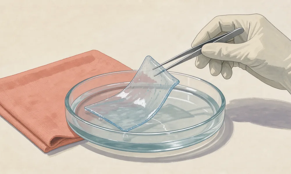
The nanoparticle and hydrogel study in mice enclosed KPV in hyaluronic-acid-functionalized particles carried within a chitosan/alginate gel. The targeted formulation reached inflamed colon tissue and reduced colitis severity more effectively than non-targeted formulations.
A later combination nanodrug study brought KPV together with the immunosuppressant FK506. In mice with acute or chronic colitis, the combination improved outcomes beyond either agent alone, restoring tight-junction proteins and lowering inflammatory cytokines. Those junctions help seal the gaps between neighboring intestinal cells.
I respect the engineering involved. The delivery material and the additional drug are active parts of the experimental question. Removing them from a summary leaves a much simpler treatment than the researchers actually tested.
Recovery in mice
The murine inflammatory bowel disease experiments reported earlier recovery, stronger regain of body weight and reduced inflammatory infiltration in the colon. Activity persisted in mice lacking MC1R, one melanocortin receptor, supporting a route that differs from the parent hormone’s usual receptor signaling.
An earlier comparison of alpha-MSH fragments in mice and cultured macrophages found another useful complication: KPV reduced accumulation of inflammatory cells but, unlike the core hormone fragments, did not suppress macrophage cytokine release in that experiment.
For me, the differences between those experiments are a reason to resist a single, all-purpose explanation. The cell type and inflammatory model affect the account of what KPV did.
Rabbit corneal repair
The rabbit corneal wound study tested topical KPV on the clear surface at the front of the eye. The epithelial surface rebuilt faster, through an effect linked to nitric oxide. This is an animal ocular-repair finding, and it supplies no established human eye treatment.
I’m satisfied to discuss the eye result on its own terms, alongside the gut experiments. Neither experiment tested a torn muscle.
Human evidence and status
The sources here contain no published human KPV treatment trial and no organized body of user reports from which benefits or harms could be counted. Human efficacy, long-term safety, interactions and pharmacokinetics remain unestablished. Small size and a relationship to a natural hormone leave those questions open.
KPV has no approved drug or dietary-supplement status and is intended for laboratory research. The sport sources establish no KPV-specific classification by name; broader non-approved-substance rules may still matter. The current answer must come from the relevant anti-doping body.
The contact-sports FAQ addresses the sports-injury claims directly. The comparison of outcomes shows how far the intestinal experiments sit from the other subjects studied here.